


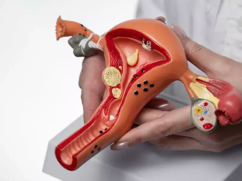
A uterine imaging test, medically known as hysterosalpingography (HSG), is a procedure in which X-ray images of the uterus and fallopian tubes are taken. During this procedure, a radiopaque fluid (a substance that makes structures visible on X-ray) is introduced into the uterus, allowing X-ray images of the uterus and fallopian tubes to be obtained. These images play a very important role in diagnosing problems related to female reproductive health.
Hysterosalpingography is primarily used to check whether the fallopian tubes are open and to detect abnormalities inside the uterus. This procedure is performed in cases such as diagnosing infertility, identifying blocked fallopian tubes, and determining structural abnormalities of the uterus.
The images obtained as a result of the uterine imaging test are carefully examined by physicians and are used to diagnose reproductive health problems and determine appropriate treatment methods. In this way, issues affecting fertility can be detected early and the necessary treatment process can be initiated.
Why Is a Uterine Imaging Test Performed?
A uterine imaging test is performed to diagnose various problems related to female reproductive health. Some of the reasons for performing this test include:
Diagnosis of Infertility Problems:
Hysterosalpingography plays an important role in diagnosing infertility. It is used to identify factors that may cause infertility, such as blocked fallopian tubes or structural abnormalities within the uterus.
Evaluation of Tubal Patency:
The test is performed to check whether the fallopian tubes are open. Blocked tubes are one of the most common causes of infertility and can be detected with a uterine imaging test.
Detection of Intrauterine Abnormalities:
Hysterosalpingography is used to identify abnormalities such as uterine polyps, fibroids, or adhesions. These intrauterine problems may negatively affect pregnancy and childbirth.
Assessment After Previous Surgical Procedures:
Previous surgical interventions involving the uterus or fallopian tubes may cause structural changes. A uterine imaging test plays an important role in detecting such changes.
Investigation of Painful Menstrual Cycles:
Some women experience painful menstrual cycles. Hysterosalpingography may be used to investigate the underlying causes of such issues.
Performing a uterine imaging test allows for early diagnosis of many reproductive health problems and helps determine appropriate treatment options.
How Is a Uterine Imaging Test Performed?
A uterine imaging test is usually performed by a gynecology specialist. The steps of the procedure are as follows:
Preparation Stage:
Before the procedure, the patient is informed in detail and an informed consent form is signed. Taking the menstrual cycle into consideration, the test is usually performed within the first 10 days of the menstrual period.
Administration of Intrauterine Contrast Fluid:
During the procedure, the patient is positioned on a gynecological examination table with her legs apart. A radiopaque fluid is then introduced into the uterus using a catheter.
Obtaining X-ray Images:
The injected fluid allows X-ray images of the uterus and fallopian tubes to be captured. The pelvic area is imaged using an X-ray device.
Evaluation and Reporting:
The obtained images are carefully examined by the gynecologist. The structure of the uterus and fallopian tubes, blockages, and abnormalities are evaluated. The results are then presented to the patient in the form of a detailed report.
The uterine imaging test usually takes approximately 30–45 minutes. During the procedure, patients may experience mild pain or discomfort. However, hysterosalpingography is generally considered a safe and minimally uncomfortable procedure.
Preparation Before a Uterine Imaging Test
To ensure the successful completion of the uterine imaging test, certain preparation steps must be followed beforehand:
Information and Consent Form:
Before the procedure, detailed information about the test is provided to the patient, questions are answered, and an informed consent form is signed.
Consideration of the Menstrual Cycle:
The test is usually performed within the first 10 days of the menstrual cycle to obtain clearer images of intrauterine structures.
Use of Pain Relievers:
Since mild pain or discomfort may be felt during the procedure, taking a pain reliever approximately 30–60 minutes beforehand is recommended.
Prevention of Infection Risk:
The patient’s risk of infection is evaluated prior to the procedure. In some cases, antibiotic treatment may be initiated.
Discontinuation of Contraceptive Use:
If the patient is using any contraceptive method, it should be discontinued at least one week before the procedure.
Bowel Preparation:
In some cases, bowel cleansing may be required before the test to ensure clearer imaging.
Preparation on the Day of the Procedure:
On the day of the test, the patient is advised to wear comfortable clothing and have a companion accompany her if possible.
These preparation steps help ensure that the procedure is performed more safely and comfortably.
What Happens During the Uterine Imaging Test?
A uterine imaging test is performed by a gynecology specialist. The patient’s experience during the procedure can be summarized as follows:
Gynecological Examination:
Before the procedure, a gynecological examination is performed. The doctor examines the uterus and fallopian tubes.
Administration of Contrast Fluid:
A radiopaque fluid is then injected into the uterus through a catheter.
Obtaining X-ray Images:
The injected fluid allows X-ray images of the uterus and fallopian tubes to be captured using an X-ray device.
Mild Pain or Discomfort:
During the procedure, patients may feel mild pain or discomfort. This sensation is usually short-lived and tolerable.
Follow-Up and Evaluation of Results:
After the procedure, the patient is allowed to rest briefly. The images are then evaluated by the gynecologist, and the results are explained to the patient.
Overall, hysterosalpingography is a safe and minimally uncomfortable procedure. However, in rare cases, complications such as infection, severe pain, or discomfort may occur. In such situations, additional treatment may be applied in accordance with the doctor’s recommendations.
The information provided here is for informational purposes only and should not be considered as medical advice. Please consult with your doctor for personalized treatment recommendations and professional medical guidance.